Parents & Families
Finding out that your child has a diagnosis of Trisomy 18 is life-changing. You face an uncertain future and have many, many questions.
It is very likely that prior to learning of your child’s diagnosis, you were unfamiliar with Trisomy 18. While considered rare medically, Trisomy 18 is a life-threatening genetic disorder that impacts about 1 out of every 2000 pregnancies in the U.S. You and your child are not alone.
We have the resources and answers you need to understand your child’s diagnosis and make informed decisions about their health. We can also connect you with other families in our community who have faced these same challenges.
What is Trisomy 18?
Trisomy 18, also known as Edwards syndrome, is a condition that is caused by an error in cell division, known as meiotic disjunction. When this happens, instead of the normal pair, an extra chromosome 18 (a triple) results in the developing baby and disrupts the normal pattern of development in significant ways that can be life-threatening, even before birth. A Trisomy 18 error occurs in about 1 out of every 2000 pregnancies in the United States and 1 in 6000 live births. The number of total births is much higher because it includes significant numbers of stillbirths that occur in the 2nd and 3rd trimesters of pregnancy.
Unlike Down syndrome, or Trisomy 21, which also is caused by an extra chromosome, the developmental issues caused by Trisomy 18 are associated with medical complications that are more potentially life-threatening in the early months and years of life. Studies have shown that only 50% of babies who are carried to term will be born alive. At birth, intensive care admissions in Neonatal Intensive Care Units (NICU’s) are routine for infants with Trisomy 18.
How Is Trisomy 18 Diagnosed?
Most cases of Trisomy 18 are diagnosed prenatally in the United States. Regardless of whether the diagnosis is made prenatally or postnatally (after birth), the process is the same. A sample of the baby’s DNA is extracted from a blood sample or other bodily cells or tissue and is cultured to examine a picture of the chromosomes called a karyotype.
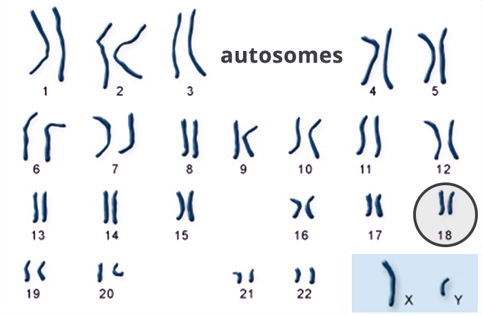
A karyotype is simply a picture of a person’s chromosomes. In order to get this picture, the chromosomes are isolated, stained, and examined under the microscope. Most often, this is done using the chromosomes in the white blood cells. A picture of the chromosomes is taken through the microscope. A visible extra 18th chromosome confirms a Trisomy 18 diagnosis.
A lot of prenatal testing is available which may indicate Trisomy 18. It is important to understand that there are two types of testing: screening and diagnostic.